Retina and Optic Nerve Imaging
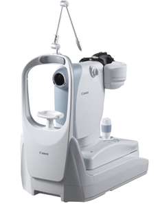
· Retinal imaging is an important tool for the screening and diagnosis of eye problems, such a retinal detachment, glaucoma, optic nerve swelling, retinal vascular disorders, hypertensive and diabetic retinopathy and macula degeneration.
· The photograph takes a few seconds on each eye and is painless.
· We will store your photograph for future comparison and to monitor for progression or regression.
· You will be able to see high resolution images of your own optic nerves, maculae, and retinal arteries and veins.
By examining the retinal vasculature, we can also get a good indication of your general health and whether certain diseases like diabetes, hypertension and hypercholesteremia are under good control.
And the best thing of all is......we do not need to dilate your eyes to do this! ***
***Note: Although this new retinal camera allows us to see very detailed high resolution images of your retina, it does not replace the need for a dilated retinal exam under certain circumstances. The doctor will let you know whether a dilated retinal exam is required or not.
Advanced Automated Perimeter
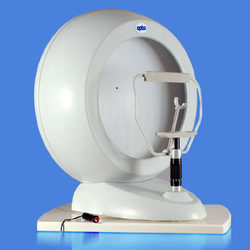
Automated visual fields are the only way to document functional visual loss and whether such loss is progressing or remaining stable over time. As such, they play an indispensable role in ocular pathologies such as glaucoma, brain tumor, multiple sclerosis, vascular problems, congenital defects, infections, or retinal diseases such as macular degeneration, retinal detachment, or inflammation.
We have always had a screening automated perimeter in the practice which is great at detecting early visual field defects. However, with this newer and more advanced perimeter, we can now more effectively follow these defects over time to determine whether there is progression or regression of the disease process.
We have always had a screening automated perimeter in the practice which is great at detecting early visual field defects. However, with this newer and more advanced perimeter, we can now more effectively follow these defects over time to determine whether there is progression or regression of the disease process.